How. Details: Measuring cells 4. To accurately measure the size of cellular structures we need a suitable scale: 5. The graticule a more suitable Using a microscope to measure cell size - The key features. How. Details: The calibrated eyepiece graticule can be used to make measurement of
Bacteria are single-celled and are classified under the domain Prokaryota. As such, they lack membrane-bound organelles like those found in eukaryotes. Although they are all microscopic organisms that can be found in various environments in nature, bacteria widely vary in size,
Estimate how many cells laid end to end it would take to equal the diameter of the field of view. You can use an ocular micrometer to measure cell size. How to Calculate Magnification on a Light Microscope. How to Estimate the Size of a Specimen With a Microscope.
Estimating Size Under a Microscope. There is two major process; First one is to measure of view field, second is to count of the You can estimate anything you want to measure in this range, based on how much of the field it covers. If you divide the length of diameter to the number of the cells
Measuring items under a microscope at low power is not difficult if you are prepared to work with Estimating Size Under a Microscope. 1. Begin sizing the microscope's viewing field at 40 times Divide the number of cells in view with the diameter of the field of view to figure the estimated
electron components sensors brookhaven synoptic milestone chemical imaging particle eliminated
Ever since scientists discovered cells under the microscope more than 350 years ago, they have noted that each type of cell has a characteristic size. From tiny bacteria to inches-long neurons, size matters for how cells work. The question of how these building blocks of life regulate their own
Micrometry (micro: microscopic, metry: measurement) is the measurement of the dimensions of microscopic objects in terms of length, breadth, diameter and Microorganisms are microscopic objects, as they are not visible to naked eye and can only be observed under microscope.
Under the microscope, animal cells appear different based on the type of the cell. Under direct observation, only the shape and size of the cell are visible because the cells are transparent and colorless. Through this technique, it is even possible to measure the length of the DNA strands.
When viewed under light microscope, most bacteria appear in variations of three major shapes: the rod (bacillus), the sphere (coccus) and Due to the presence of a rigid cell wall, bacteria maintain a definite shape, though they vary as shape, size and structure. When viewed under light
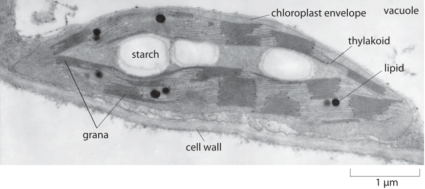
chloroplasts chloroplast electron micrograph cell grana thylakoid membrane figure biology stacks per
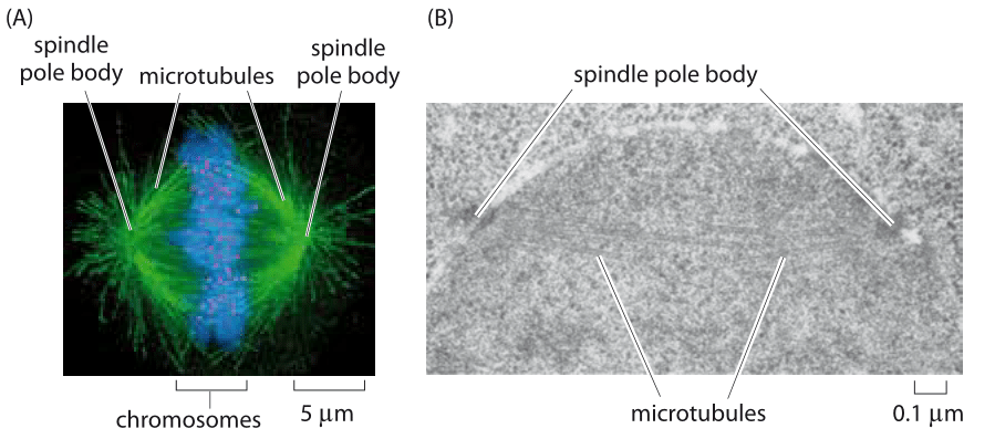
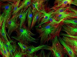
cell microtubules filaments microtubule spindle cytoskeleton hela cells distribution human mitotic actin fluorescence
In order to estimate cell size during microscope investigations, the microscope should be calibrated. This means using a stage micrometer (a microscopic ruler), a calibration grid to measure the distance across each field of view (FOV1). As you increase the magnification the FOV decreases.
> It is possible to use a linear scale inserted into the eyepiece objective lens (or use a special eyepiece lens that contains a scale) to measure cell length (or anything that can be clearly seen under the microscope). > However, the eyepiece scale (graticule needs to be calibrated before
key
Learn about the microscope's field of view and how to calculate using a formula from our experts at Say, for example, you are viewing a cell or specimen under an optical microscope. A crude way of measuring the field of view is by using a ruler under the microscope for a particular magnification.
To measure the size of the specimen under the microscope, the ocular micrometer is used. How to Use an Ocular Micrometer. The magnified image of the specimen is formed on the ocular micrometer and that the micrometer scale and the sample image can be viewed simultaneously through
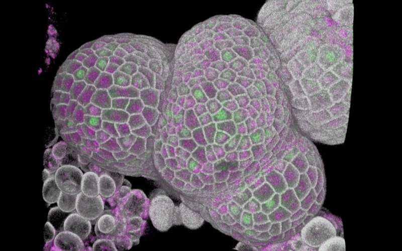
How can you see a plant cell under a compound microscope? Each green spot is a particular six base sequence (restriction site), labeled via a few steps. By measuring the distance between these sites a map can be built which can be used to check and improve the determined sequence of
Linear Measurements (Micrometry). The first reported measurements performed with an optical microscope were undertaken in the late 1600s by the Dutch Alternative mechanisms for performing measurements at high magnifications in compound optical microscopy must be employed, and
put ruller under the MS and mesure the diameter rember 1 milliter equals 1000 micrometers so if the diameter which im guess you no what that is say its millimeter times that by thousand and it become 2700 microns now you count how many cells fit in that dimater to get the approximate lenght so if 6
cells microscope skin under human
How can it bee seen? Electron microscope (EM) Light microscope or EM. How big are the objects you've just measured? Use your class resources to approximate their. Scale: The relative size of an object Size: The measurement of how big (or small) an object is.
innes
We can measure cells then make comparisons between different types of cells. After determining field sizes, give sizes of some organisms and have students decide which microscope Ask students how speed of movement or interactions with other organisms would influence their choice
Materials. Microscope. Ocular micrometer. Place a stage micrometer on the microscope stage, and using the lowest magnification (4X), focus on the grid of the stage micrometer. Since each division of the stage micrometer measures 10 micrometers, and since you know how many ocular divisions
If this was helpful, please is a short video on how to find the size of an object when looking at it through a microscope. Size of

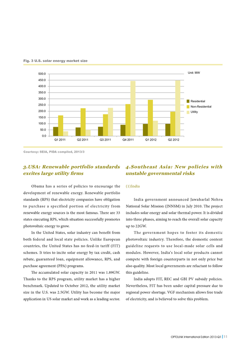
pida key
How does a microscope work? How can we measure the size of a cell? What is the advantage of studying structures with a microscope? Light microscopes using visible light and lenses to form a magnified image of the object under investigation cells of plant or animal tissue.
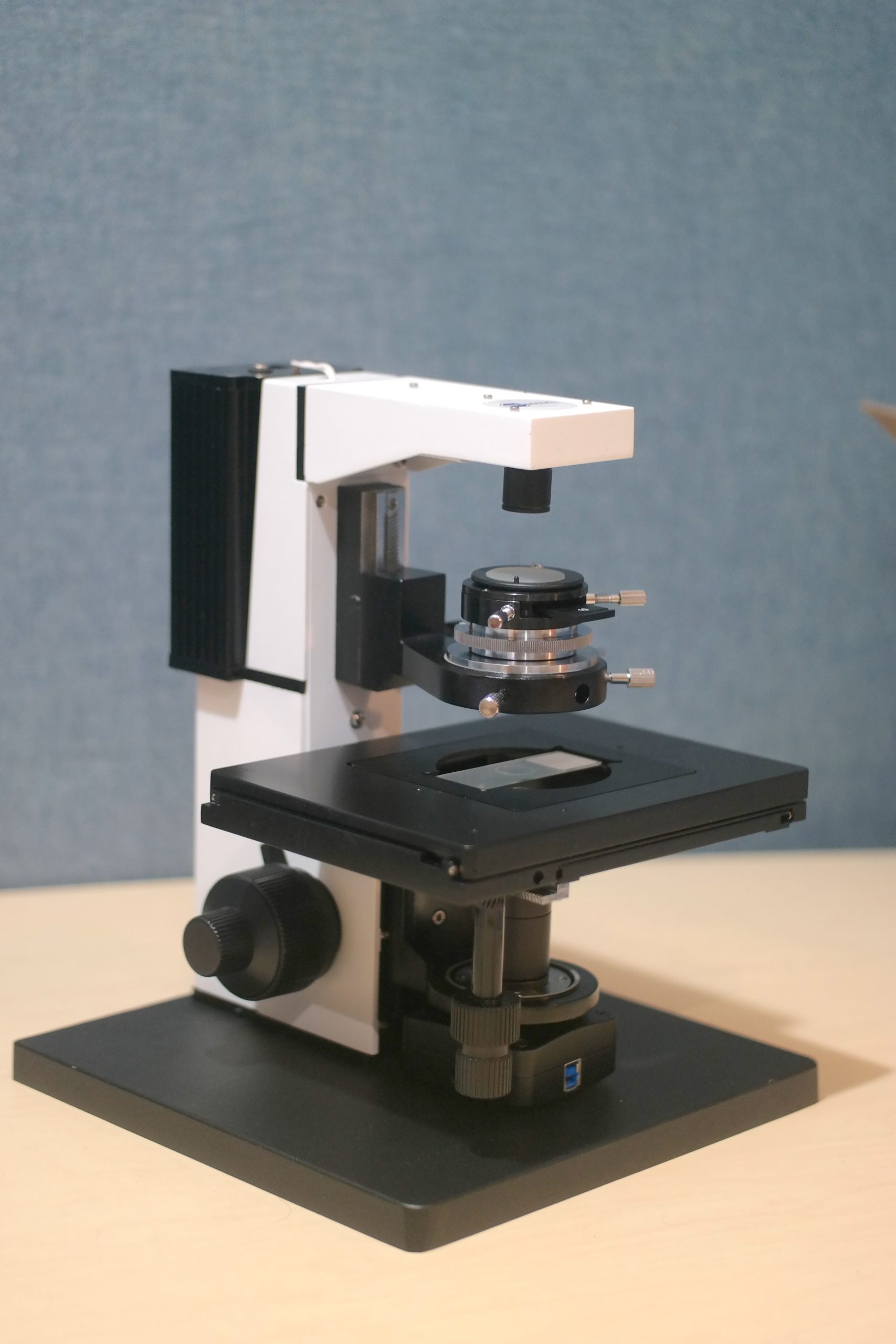
microscope incubator is220 bioimager
Measuring with the microscope is simple when you understand a few steps to take before using your microscope eyepiece reticle. When looking through the microscope, the reticle image is imposed upon your specimen image. Most often the reticle is used to make measurements or count particles.
Contents 13 How are microorganisms measured under a microscope? 14 How does Imagej measure cell size? Two types of electron microscopy—transmission and scanning—are widely used to study cells.
Since objects under the microscopic are usually too small to measure using an ordinary ruler, we must use more intricate and mathematical methods to There are two predominant ways to measure specimen size under a microscope if you don't have specialized microscope camera software.
Estimating the Size of Cells Using a Compound Light Microscope. 1. Learn how to use the compound light microscope. 2. Learn how to make a preparation for viewing on a slide. 6. Draw a bar graph comparing the lengths of the various cells you measure.
How the size of individual cell can be measured by a typical SSC vs FSC graph without using a reference (known size beads or cells)? Although from a typical SSC vs FSC graph, we can find out relative size and complexity of the cells belonging to a particular population, how can we
This video covers how to measure objects viewed through a light microscope in 10 practical video on how to measure cells on a micrograph as well as converting from micrometres to millimetres. 082 - How to look at Potato starch grains under the microscope | Microscopy.
Measurement with the Light Microscope. Your microscope may be equipped with a scale (called a Be aware that even under the best of circumstances the limit of resolution of your microscope is 1 or 2 For example, suppose you measure the length of a flagellum on a Chlamydomonas cell at
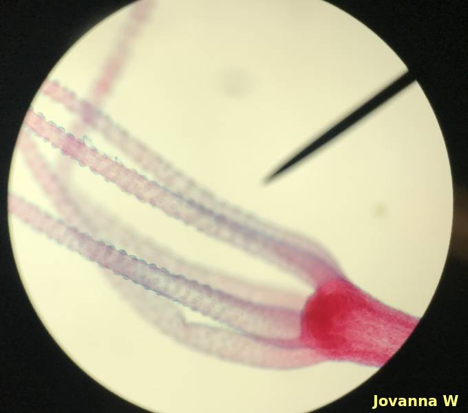
microscope hydra letter 400x power specimen field estimate lab across diameter nicerweb locked threads fits times
Cell size can be measured using an eyepiece graticule. The graticule has a ruler on it. You must find out the distance measured for each division of the graticule. The calibrated eyepiece graticule can be used to make measurement of any cells or other structures viewed with the microscope on
We measured the cell size by using a microscopic meter slide. You can directly observe the mold under a stereo microscope or prepare a spore specimen for a compound microscope. When you look for stuff to see under your microscope, do not forget all kinds of everyday things in your kitchen!
