
ultrasound uterus normal ovaries
17, 2018 · Ultrasound scans use high frequency sound waves to capture images and video of the inside of the body. Abdominal ultrasounds to help your doctor see the organs and structures inside the abdomen. ...

weeks ultrasound days
We have created this ultrasound image optimization guide to provide you with a plain-English Knowing they exist is one thing, but learning how to use them is a very important step in getting With Edge Enhancement, the ultrasound attempts to make a sharper image by combining adjacent signals.

ultrasound twins week triplets
An ultrasound is the most common way to determine a baby's gender. What differences can be seen between boy and girl ultrasound pictures? If it is somewhere in between, it may be harder to make a definitive determination. Flow of urine: The flow of urine can sometimes be spotted in a fetus.
’s Picture Archive and Communication System (PACS) makes it easy to see your patient’s reports and images immediately after the imaging exam is performed and the report is finalized. Additionally, you will have historical access, with approval, to your patient’s imaging performed through ARA or one of our partners for comparison purposes.
I intend to make this a large library of ultrasound images obtained from friends in the medical world. 2) Each ultrasound image is fine tuned; , every effort is made to adjust contrast, brightness and sharpness (among many parameters) to bring out the best in each picture.
If you have had an ultrasound imaging done on your or any of your close ones recently and want to know how to interpret the images yourself without taking the help of a doctor, you first To help you out in your endeavor, we have presented this complete guide on how to read an ultrasound picture.

funny ultrasound things detected sound ultra even famous born klyker creepy random internet days before comment viraluck
Ultrasound (sonography, Doppler study) is a non-invasive diagnostic medical procedure that examines internal organs and various parts of the body. What Facts Should I Know about Ultrasound? Ultrasound is a diagnostic or screening tool to confirm medical disorders or to assist in
A sonogram or ultrasound picture is a black and white photograph, which can be a little confusing to interpret at first. Once you appreciate ... read more. I went to an outside Ultrasound place at 15 weeks 2 days. The tech took a while to get the baby to move its legs out of the way, the baby w ... read more.
An ultrasound may be performed for a variety of reasons, but looking at a baby in the womb is the If you have recently had an ultrasound and you want to know how to interpret the images on your We use cookies to make wikiHow great. By using our site, you agree to our cookie Settings.
second-trimester anatomy ultrasound, or structural survey, can be done at 18-20 weeks. Gender determination upon request. This is the ultrasound that your doctor will send you to make sure all the organs inside your baby are developing fine. This is also when you find out whether you are having a boy or a girl.

choledocholithiasis radiopaedia duct modality bile uterus gallstone sonography
Getting problematic on how to make a fake ultrasound picture with how busy you are? Well, it's about time not to overcomplicate things! offers the fake 2D ultrasound and 3D ultrasound, and you could get these fake ultrasounds as is, or with personalization.
Ultrasound Courses, Registry Review and CME Training Products for Cardiology, Vascular Surgery, Radiology Physicians, NP’s, PA’s, Sonographers, Military, and other Medical Professionals. Gulfcoast Ultrasound Institute is the most flexible and proven ACCME Accredited Ultrasound CME in the industry.

sarcoidosis liver granuloma case findings caseating non liveratlas
How to Deal With Bullies: A Guide for Parents. Mean kids aren't just a middle-school problem. The trouble has trickled to the youngest grades. An early pregnancy ultrasound may be done transgvaginally so doctors get a clearer picture of your baby. In this case, the OB-GYN will place
Ultrasound uses sound waves you can't hear. When a provider glides the probe over a special gel applied to the testing area, the device captures pictures of soft tissues inside Gives you instructions: The professional performing this test has received training in how to achieve the clearest images.
Sound waves will make pictures just when they can reflect from a surface. For instance, sound waves travel effectively through liquids and won't ricochet off of water How Does an Ultrasound Work? As a rule, an ultrasound machine estimates sound waves that have bobbed off the body's inward tissues.
Browse royalty-free stock video for Ultrasound in popular formats including 4K and Full HD for your creative needs. Download your videos today!
Why Ultrasound? Over half a century old technique! Arguably the most widely used imaging technologies in medicine. Portable, free of radiation risk, and relatively inexpensive compared to MRI, CT and PET Tomographic, , offering a "cross-sectional" view of anatomical structures.
We also describe how to stream a picture-in-picture ultrasound feed during a videoconference. Point of care ultrasound is important to the specialty of Physical Medicine and Rehabilitation (PM Based on the voter output, a majority rule is then used to make the final decision of the active talker'
Ultrasound imaging uses sound waves to produce pictures of the inside of the body. It helps diagnose the causes of pain, swelling and infection in the body's internal organs and to examine an unborn child (fetus) in pregnant women. In infants, doctors commonly use ultrasound to evaluate the brain,

ultrasound ovaries polycystic
Ultrasound is sound waves with frequencies higher than the upper audible limit of human hearing. Ultrasound is not different from "normal" (audible) sound in its physical properties, except that humans cannot hear it.
While little hard evidence on how to improve 3D ultrasound pictures exists, doctors who conduct ultrasounds have found certain lifestyle changes may improve images. You want to make sure you get your ultrasound in during a time in pregnancy where good pictures are more likely.
How to Make a Fake Ultrasound Picture - Make A Fake Ultrasound Picture - How. Details: Download Ultrasound Prank Free - Pregnant Spoof And Fake Pregnancy Trick and enjoy it on your iPhone, iPad
See more ideas about ultrasound pictures, new baby products, ultrasound. This is a print from my original illustration . The image is centered to fit with the paper size leaving a white border around. All my Prints are made by me, in a lovely heavyweight Epson paper 200g/m2 .
06, 2020 · Fetal ultrasound: A fetal ultrasound, or sonogram, is an imaging technique that uses high-frequency sound waves to produce images of a baby in the uterus.

kidney sponge medullary
How do patients prepare for an ultrasound? How are the results of ultrasound interpreted and Ultrasound produces sound waves that are beamed into the body causing return echoes that are The ability to measure different echoes reflected from a variety of tissues allows a shadow picture
04, 2022 · For example, the 4D ultrasound is an optional scan since a 3D ultrasound usually suffices to evaluate the baby. Unfortunately, it is illegal in some countries due to various reasons. Read on to learn more about the 4D ultrasound scan, how it works, what you can expect, and whether your insurance covers it.
We all know that some ultrasound photos fade which is horrible because we all want to keep that moment and make it last I am the same way. I had my son's ultrasound photo up on my mirror for a long time (okay he is two right now so a year or so) until I noticed that my mom's
How does an ultrasound work? Ultrasounds use high frequency sound waves to create pictures of your fetus and reproductive organs (like your uterus and cervix). An ultrasound — also called a sonogram — helps your doctor look at your fetus to make sure it's developing normally.
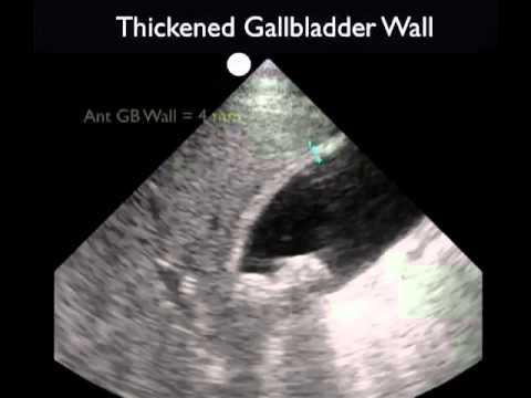
cholecystitis ultrasound gallblader sonosite inc sono gallbladder sonography anatomy
How is ultrasound made? It's impossible for us to make ultrasound the same way we make normal sounds—by hitting and blowing things, as Photo: This pregnant woman is watching an ultrasound scan of the baby developing in her womb. Note the ultrasound scanner (bottom right) being
Ultrasound is a non-invasive, immediate tool used to image tissue. It will not penetrate bone (like an X-Ray). So the first step to help you read the ultrasound image is to be familiar with the anatomy that you are imaging. Various body tissues conduct sound differently. Some tissues absorb sound
Next let's go over how an ultrasound device uses ultrasonic waves to create pictures on the screen for you. Understanding how these waves behave will be helpful in understanding how to optimize your ultrasound settings and images. I'll make it as simple as possible for you and just go over
07, 2021 · Sometimes 3D or even 4D ultrasound technology is used instead of 2D. To get the most comprehensive anatomy assessment, the sonographer will be aiming for many different views from lots of different angles. When the technician gets a clear shot, they will freeze-frame the picture (that's the actual sonogram) and measure a specific part of the body.
How to Make a Fake Picture With Famous People. Is She Faking a Pregnancy or Is the Baby Real? Question by Walking Contradiction: I have put a Technorati Tags: Baby Ultrasound Picture, make a ultrasound picture, make your own ultrasound,. Can you make fake ultrasound pictures?
© 2016 Conquest Imaging. How Do We Generate an Ultrasound Wave? Ø Tissue absorption of sound energy contributes most to the attenuation of an ultrasound wave in tissues. Recent advancements in computer technology and software engineering make 4D ultrasound imaging Picture Archive and Communications Systems - DICOM Medical imaging storage server that
17, 2020 · To read an ultrasound picture, look for white spots on the image to see solid tissues, like bones, and dark spots on the image to see fluid-filled tissues, like the amniotic fluid in the uterus. If you're 12 weeks along in the pregnancy, you may be able to make out your baby's head, and if you're 20 weeks along, you may even see the spine ...
Ultrasound, also called sonography, uses sound waves to develop ultrasound images of what's going on inside the body. An instrument called a transducer Ultrasound technicians, or sonographers, have special training in how to perform the test. Then a radiologist or your doctor will interpret
Baby Ultrasound 5th Month - Boy Baby. fetal ultrasound of 16 weeks 17 weeks baby boy moving.
By sharing your ultrasound pictures with the subreddit, which means we're able to share the most useful information we can about the process. For instance, we can find out the exact moment of your delivery, and the exact day of your surgery. With that information, you can tailor your
