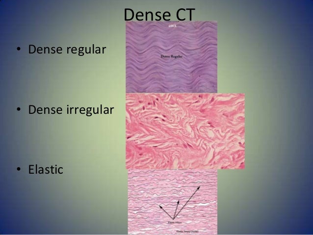
connective irregular areolar reticular cartilage adipose hyaline
Microscopes are generally made up of structural parts for holding and supporting the microscope and its components and the optical parts which are used for magnification and viewing of the specimen images. This description defines the parts of a microscope and the functions they perform to
Thank you for visiting microscope slide label templates page! Sit back and take a look at our vibrant range of carefully-created top quality templates. In case you presume it's going to cost the earth, TemplateMonster will bust this myth. We wager our coders have already built a skin that is
neuron multipolar motor cell dendrites axon embed gettyimages

When preparing microscope slides for observation, it is important first to have all necessary materials on hand. This includes slides, cover slips, droppers or pipets and any There are two different types of microscope slides in general use. The common flat glass slide, and the depression or well slide.
The microscope uses bright light to illuminate through the specimen and provides an inverted image at high magnification and resolution. There are two lenses that magnify the image of the specimen - the objective lens on the nosepiece and the ocular lens (or eyepiece).
Microscope slides can be prepared with a dry mount, a wet mount, and a prepared mount. Each type of mount is used depending on the sample that will be examined and observed under the microscope. Each type will also require specific skills and knowledge before it can be performed as there
Bulk Sample Microscope slide Preparation Step by step guide shows how to prepare slides of bulk sample materials for forensic analysis - Lab SOP. POST a QUESTION or COMMENT about how to prepare a bulk sample of , scraping, or other large particles for successful forensic analysis
The google slides shown below have the same microscope image with the labels for students to copy. Introduction to the Light Microscope - students focus the letter "e" on prepared slides and Investigation: How Can a Microscope be Used to Make Observations - this activity is designed
How do stereo microscopes work? The difference between Compound and Stereo (Dissecting) How do stereo microscopes work? The working principle of Stereomicroscopy depends on two light 3. Switch on the light sources. For microscope slides or other transparent objects, bottom
may use your textbook and class notes to help you identify the stages of mitosis as seen under the microscope. Purpose: The student will correctly identify and draw four stages of mitosis using microscope slide images of onion root tips and whitefish blastulae. Procedure: The slides below show longitudinal sections of allium (onion) root tip.

microscope parts printable learning lesson plans

histology vein lining blood vessel epithelium squamous simple epithelia endothelium drawing venule features stain cardiovascular covering system skin vessels epithelial
Microscope labeled diagram. 1. The Microscope Image courtesy of: Basic rules to using the microscope 1. You should always carry a microscope with two hands, one on the arm and the other under Clipping is a handy way to collect important slides you want to go back to later.
Microscope Slide Preparation. Microscope slides are pieces of clear glass or plastic that hold a sample and allow it to be viewed through a light microscope. 3. Make two smears, let them air dry, and label them clearly. Place the dried slides in the slide transport containers provided.
Drawings. When drawing what you see under the microscope, follow the format shown below. It is important to include a figure label and a subject title above the image. The species name (and common name if there is one) and the magnification at which you were viewing the object should be written below the image.
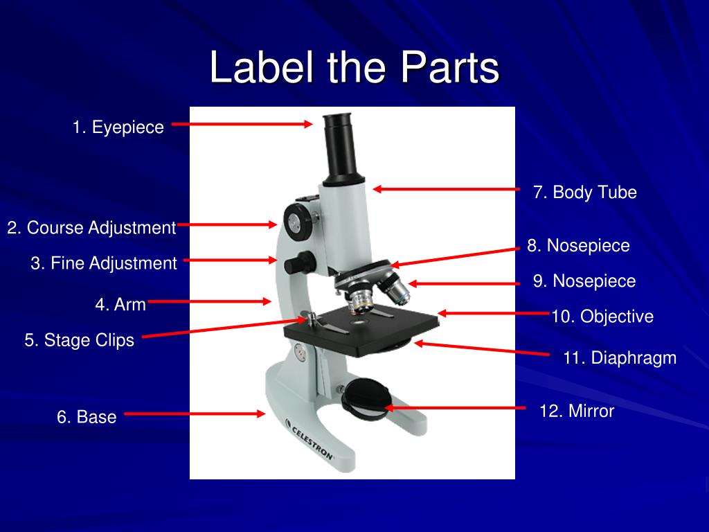
parts microscope functions label ppt tube objective slide course adjustment arm which amount fine lower eyepiece through powerpoint presentation nosepiece
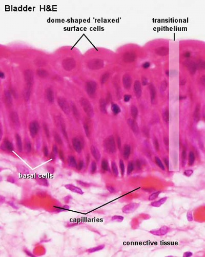
histology urinary bladder epithelium transitional lining system epithelia skin cells tissue epithelial urine pseudostratified urethra ureter embryology covering simple ureters
Slide labels will help you effectively id and keep track of samples. These labels have a strong self-sticking adhesive that will stay in place even if they are stored Slide labels resist chemicals and will not smear, fade or fall off when exposed to chemicals and solvents typically used in the lab for cleaning.
You make your own homemade microscope slides! I had already swapped some Virtual Assistant work for the microscope so that was a big win for That's way out of our current budget so I began to Google how to make your own microscope slides. I came up with a handful of decent websites
There are different kinds of microscope slides and cover glasses and in this video I want to give you an overview of them. Here I show you how you can make nice printed labels for permanent microscope slides.
provides a comprehensive range of quality cryostats, products and services for your histology, Mohs, and on-site pathology labs.
on the pond, students will be able to observe a variety of living things under the microscope. While students will be able to identify animal organisms under the microscope from the fact that they move, they may have some difficulty identifying some organisms, which may look like plants or fungi.
Slide Labels. How to Choose the Right Label, Ribbon, Hardware and Software for Labeling Your Slides. Some commonly used label materials include: Duraslide superior chemical-resistant microscope slide label/ribbon kits that withstand abrasion and exposure to acetone, ethanol,
Buy stereo, digital and compound microscopes from Dino-Lite, Meiji Techno, Motic and Omano. 1-877-409-3556 support@ When contacting us your privacy will be strictly protected and you will not be required to provide any personal information.
Copy of Label a microscope Labelled diagram. Microscope slide - label the parts Labelled diagram. how to use a light microscope Rank order.
2 labeled images and slides—are you labeling the right structure Completely labeled images and slides. Everything in blue must be labeled. Total magnification. Remember you multiply the ocular lens (10x) x the objective lens for the total magnification. Image 1: …
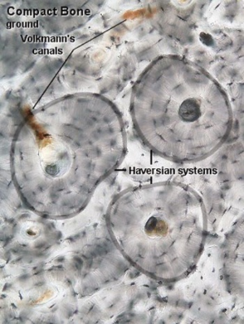
histology bone haversian lacunae system bones section canaliculi anatomy draw lamellae compact canal matrix osteocytes magnification drawing block cells physiology
How the specimen slide must be prepared largely depends on its properties, such as whether it's organic or inorganic, live or fixed, wet or dry, thick or thin, large or small, and so on. In fact, these things determine not only how the specimen preparation will go, but also the type of microscope that
tissue histology connective cell cells dense collagen mesenchymal quiz derived histamine which material
Microscope 101: How To Properly Prepare a Microscope Slide. Often times, new microscope users are puzzled by how to properly prepare slides. A slide incorrectly prepared can often lead the novice microscopist to believe their new microscope is defective (as we're more prone to think
material from Section of your text to label the condenser, objective, and ocular lenses in the diagram of the compound light microscope in Figure 1. Describe the function of each lens in producing the magnified image of a specimen: Condenser Lens: (#4) gathers light from light source and allows a small cone of light to reach a portion of
each slide and view them one at a time with your microscope experimenting with different magnification. Write down your observations about each to see how hairs from humans and animals differ. You can also look at threads or fibers from …
Microscope slides are your gateway into a whole new world. Learn about the different types of slides and how to prepare them here. Microscope slides are amazing little things. With a few simple steps, prepared slides can catapult you into a whole world that you never realized existed.
A microscope slide is a thin flat piece of glass, typically 75 by 26 mm (3 by 1 inches) and about 1 mm thick, used to hold objects for examination under a microscope. Typically the object is mounted (secured) on the slide, and then both are inserted together in the microscope for viewing.
Make sure to know how to label objective lenses, stage clip, stage control, coarse adjustment, fine adjustment, voltage dial. Only RUB Labeled Microscope & Quiz 2 Study Guide. How would you move the stage side-to-side so that the slide is centered under the objective lens?
investigators use elaborate microscope slides called McMaster slides. These are chambered with a grid pattern overlay and will provide a more scientific and precise value of the infestation but you do not need these special slides to determine if there is a problem or not. ... Be sure to label the container with the date, time and animal ...
01, 2022 · If you have microscope slides, these are perfect to use for putting a fingerprint on. If not, any smooth table, chair, appliance, wall, floor, doorknob, or faucet will do. Advertisement. Part 2. Part 2 of 2: Gathering the Prints Download Article 1. Press your finger (or fingers) hard on the smooth surface. ...
How to create your own microscope slides and ideas of different samples to view under the microscope. Creating permanent slides involves sealing your specimen within the slide so it lasts, and then labeling the slide so that years from now you remember what the sample was.
A microscope is one of the invaluable tools in the laboratory setting. It is used to observe things that cannot be seen by the naked eye. Microscopes are specially created to magnify the image of the subject being studied. This exercise is created to be used in homes and schools. the
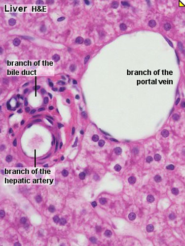
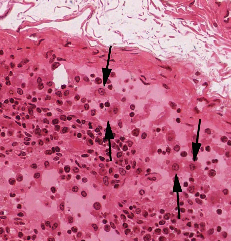
liver histology bile portal human slides tract system slide triad duct endocrine digestive anatomy tissue identify gallbladder macrophages pancreas sinusoids
Microscope slides are pieces of transparent glass or plastic that support a sample so that they can be viewed using a light microscope. There are different types of microscopes and also different types of samples, so there is more than one way to prepare a microscope slide. The method used to
Microscope slides are made of glass or plastic, approximately 1x3 inches and between mm thick. Multiple methods of preparation allow for advanced viewing of Objects magnified under compound microscopes are mounted onto microscope slides. Made of glass or plastic, slides
Microscope slides are used to examine single-celled organisms and to look up-close at small plants and organisms. There are two types of prepared slides: dry mounts and wet mounts. Each type of preparation method is used for
Label each slide and view them one at a time with your microscope experimenting with different magnification. Write down your observations about each to see how hairs from humans and animals differ. You can also look at threads or fibers from furniture, rugs or clothing from around your house.
What are permanent slides: Permanent slides carry specimens that are preserved and mounted in mounting medium. They can be kept for a long time. Sometimes microscopes already come with a set of permanent slides. They allow you to observe specimens which are otherwise difficult if
optical microscope, also referred to as a light microscope, is a type of microscope that commonly uses visible light and a system of lenses to generate magnified images of small objects. Optical microscopes are the oldest design of microscope and were possibly invented in their present compound form in the 17th century. Basic optical microscopes can be very …
In this activity, students will label the parts of a microscope and explain what each part is for (if applicable). In order to study cells in detail, it is important that students learn how to use a microscope. Before your students use microscopes in the classroom, they should understand
