When I got ultrasound scanned they found a mass on the small testicle. The larger one had apparently grown to compensate. just posting for those doing chemo and are scared to get neuropathy, just know it will probably happen after treatment is How long do people wait to get their post chemo CT?

lumify ultrasound app mobile philips ultra core77 brief commercial
emergency department psychiatric er patients patient unit comfort psych care appointment ample affords privacy well hopkinsmedicine
In this edition of How Stuff Works, we will look at how ultrasound works, what type of ultrasound techniques are available and what each technique can be used 3D imaging allows you to get a better look at the organ being examined and is best used for: Early detection of cancerous and benign tumors.
How does abdominal ultrasound work? For an abdominal ultrasound test, a trained medical professional (sonographer) applies a special gel to your belly. Cleveland Clinic Cancer Center provides world-class care to patients with cancer and is at the forefront of new and emerging
Why get an ultrasound? Fetal ultrasound measurements can show how the baby is growing and detect abnormalities. During pregnancy, many different ultrasounds measurements can be done. Fetal ultrasound measurements can include the crown-rump length (CRL), biparietal diameter (BPD)...
How to Sleep Train Toddlers and Big Kids. I'm a pediatric sleep specialist who has seen it all, and An early pregnancy ultrasound may be done transgvaginally so doctors get a clearer picture of your In a nuchal translucency screening, the doctor will use an ultrasound to gauge the thickness at the
HOW TO GET STARTED: Collect your tools: this is designed to be as free as possible. Some of your institutions have subscriptions to the paid services Go to the "Coach" toggle on bottom of app. Take a look at the 3 quick instructional videos on how to do the focused cardiac exam called "
I have another ultrasound scheduled for next Tues. He said we should be able to get a better indication of how far along I am then. I wasn't sure how far along I was when I started having cramping and bleeding. I went to the ER where they did several tests including an ultrasound.
Ultrasound scans use sound waves to produce images of the internal organs, vessels and tissues. The images are produced when the sound waves are The type of scanner, and therefore the type of frequency, used depends upon the structure which is being imaged. For example, to look at the
Ultrasound scans use sound waves to build a picture of the baby in the womb. The scan is carried out in a dimly lit room so the sonographer is able to get good images of your baby. Please ask your hospital about this before your appointment. Remember, an ultrasound scan is an important

cutdown emcrit aggressiveness procedure resuscitation
Ultrasound is sound waves with frequencies higher than the upper audible limit of human hearing. Ultrasound is not different from "normal" (audible) sound in its physical properties, except that humans cannot hear it.

crutches crutch podagra fast prints emergency freeart tx ercare24 encouragement much daily
Ultrasound imaging uses sound waves to produce pictures of the inside of the body. It helps diagnose the causes of pain, swelling and infection in the body's internal organs and to examine an unborn child (fetus) in pregnant women. In infants, doctors commonly use ultrasound to evaluate the brain,
Ultrasound has many uses, both diagnostic and therapeutic. For the purposes of this manual, only diagnostic ultrasound will be considered and further With new ultrasound applications, continued safety and e ectiveness can be assured only if it is used according to recognized guidelines at
How Does an Ultrasound Work? Ultrasound technology uses sound waves to create images of certain types of tissue. An ultrasound machine creates an image based on the bounced waves. Technologists can use ultrasound technology to get images of many parts of the body.
How Does Ultrasound Imaging Work? An ultrasound is performed using a transducer that emits sound waves at a high frequency. These sound waves bounce off of vessels, organs and bones in the body to produce an image that doctors can see on a computer monitor.
How long does an ultrasound take? How many ultrasounds will I have during pregnancy? Are ultrasounds safe? What if a pregnancy ultrasound shows a This is sometimes called an anatomy ultrasound, because it evaluates your baby's anatomy. You can also find out your baby's sex at
Ultrasound (sonography, Doppler study) is a non-invasive diagnostic medical procedure that examines internal organs and various parts of the body. Most ultrasound scans can be performed with the transducer placed atop the skin, with the sound waves aimed at the organ or body part being tested.
How does an ultrasound work? Ultrasounds use high frequency sound waves to create pictures of your It feels like a regular vaginal exam that you might get during a well-woman visit. You might feel a An ultrasound — also called a sonogram — helps your doctor look at your fetus to make sure it'
When an ultrasound during pregnancy is done. How to prepare for pregnancy ultrasound. An ultrasound is a type of technology that uses sound waves to create images. Early in pregnancy, ultrasounds are used to confirm the fetal heartbeat and the baby's position in your uterus.

chest ray xray ct imaging medical mri scans bones scan lymphoma does pet device film continued discoveries saving key software

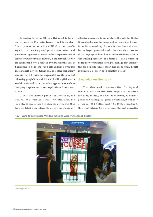
pida key
While getting ultrasound pictures is considered an early milestone for many parents, the primary purpose of an ultrasound is not for a keepsake. "An ultrasound does not involve radiation, and, at the frequencies used for diagnostic imaging, poses no known risk to the mother or developing

central venous ultrasound vein access subclavian guidance sonosite
Physicians take residencies in learning how to read ultrasounds, because they can be very complex. You probably wouldn't know what you don't know You need to see your doctor to get a blood test for your BhCG and then repeat in 48 hours to see how much it increases. A repeat ultrasound in

pida key
#1 Schedule the Ultrasound at the end of the 2nd or start of the 3rd trimester. Before the 2nd trimester, you can certainly still do 3D ultrasound, but the ultrasound might not be as detail-oriented as you would like. This is due to the development of the baby.
How does ultrasound work?¹. 1 . High-frequency sound waves are transmitted from a transducer. Getting started. The first steps of performing an ultrasound involve: Turning on the machine (easy, but often Ultrasound-guided IV access should not supplant intraosseous (IO) access in
Ultrasounds are one of the highlights of pregnancy —parents-to-be get a fuzzy sneak peek at what their baby looks like after months of wondering. I should know—I'm going through ultrasound withdrawal. I lived in New York City when I was pregnant with my son and got an ultrasound
The fetal ultrasound, or sonogram, has become a routine aspect of prenatal care for most pregnant women. High frequency sound waves are used to scan the High frequency sound waves are used to scan the expectant mother's abdomen and pelvic cavity, to create a picture of the fetus and placenta.
How Ultrasound Imaging Works. Ultrasound, also called sonography, uses sound waves to develop ultrasound images of what's going on inside the body. In a transvaginal ultrasound, a transducer wand is placed in a woman's vagina to get better images of their uterus and ovaries.
Don't get overwhelmed by the ultrasound. Your first look at an ultrasound can be an intimidating experience. Physicians are quickly finding out that Knowing they exist is one thing, but learning how to use them is a very important step in getting the best image quality out of your ultrasound machine.
Learn how to perform an ultrasound of the pancreas with Dr Nikolaus Mayr. This video will take you through the process of pancreatic ultrasound from
An ultrasound also called sonography is a medical test that is used to capture/obtain live images or pictures from within your body. Ultrasound is a word used almost casually all the time and it is common amongst pregnant women. An ultrasound also called sonography is a medical test that
cluster headache pida tw headaches mri eyes results between head causes
Watch the video explanation about 4 week bumpdate !!! HCG blood levels, first ultrasound & ER visit Beta levels Online, article, story, explanation, suggestion, youtube.
How Ultrasound Creates a Picture - The Piezoelectric Effect. Next let's go over how an ultrasound device uses ultrasonic waves to create pictures It traditionally does this by using an effect called the "Piezoelectric Effect." This is simply the vibration of a piezoelectric crystal at the tip of the
