confocal microscope schematic 3d beam imaging geometry figure lineas uv es
Confocal Microscope. High-speed acquisition of high-definition images. Users can easily learn how to control different imaging systems with a common interface and workflow. • A built-in AOTF allows fast and efficient selection of wavelengths and power modulation.
Confocal microscopes can provide high-quality scans of volumes, which nd Notice how the LFM data acquisition (bottom row) is faster both at capture time and rendering time, and requires The next step is to build a data set containing pairs of confocal stacks and corresponding LFM images.
confocal microscope computer digitizer transfers digital microscopes cas miamioh

Using a confocal microscope builds images from thin sections of a sample. By scanning several thin sections through the sample, it's possible to create a From there, you'll need to determine whether you want to use fluorescent dyes on your samples, as well as what kinds and how many different kinds.
The confocal microscope utilizes state of the art technology and lasers that separate light waves, allowing you to view images without blurred edges and in higher resolutions. In order to understand how a confocal microscope operates, an understanding of fluorescence is needed.
PDF | Confocal microscopy is a technique for increasing the contrast of microscope images, particularly in thick specimens. In this chapter, we will first discuss the principle of confocal microscopy and then the design of confocal microscopes.
15. HOW DOES A CONFOCAL MICROSCOPE WORK Confocal microscope incorporates 2 ideas : 1. Point-by-point illumination of the specimen. 2. Rejection of out of focus of light. Light source of very high intensity is used—Zirconium arc lamp in Minsky's design & laser light source in modern design. a)...

confocal microscope zeiss courtesy swiss ch

How does Confocal Microscope Work? There are some lenses inside the microscope, these By scanning many thin sections through your sample, you can build up a very clean To explain how a confocal microscope displays a depth of field, look at the imaginary cell with a imaginary organelle.

confocal lsm clsm
confocal microscope (from Noran) uses a special Acoustic Optical Deflector in place of one of the mirrors, in order to speed up the scanning. This uses a high-frequency sound wave in a special crystal to create a diffraction grating, which deflects the laser light (actually, the first diffraction peak is used, with the zeroth-order peak being thrown away).

confocal crest microscopes konfokal microscopios konfokale microscopi mikroskope
In confocal microscopy fluorescence excitation occurs throughout the depth of the tissue and out-of-focus fluorescence is rejected by the pinhole; in In 1999 I built a video-rate confocal microscope utilizing the same resonant scanner [Callamaras et al.,1999], but employing a Data Translation
Thank you for purchasing a Confocal Microscope A1. We are sure that the A1 will greatly contribute to your research with its many excellent functions. Note: When not only a confocal microscope is connected but also a camera, the Driver selection dialog box How to Use Time Measurement.
microscopy provides the ability to collect clear images from a thin section of a thick sample with low background and minimal out-of-focus interference. Optical sectioning is a common application in the biomedical sciences and has been useful for materials science as well. In practice, a sample is put on the microscope stage and an ...Author: Amicia D. ElliottPublish Year: 2020
What is a Confocal Microscope? The concept of confocal microscopy was initially developed by Marvin Minsky in the 1950s, at Harvard University with an aim of viewing the neural network without staining the tissues but it did not bear fruit due to lack of enough light source and a
The confocal microscope is so called because the illumination pinhole, the plane of focus within the specimen By restricting the illumination to a pinhole, rather than illuminating the entire field of view at once it becomes possible to build up a blur-free image in three entire image is

periscope beam microscopy uci
Introduction to Confocal Microscopy - Confocal microscopy offers several advantages over conventional widefield optical microscopy, including the ability to control depth of field, elimination or reduction of background information away from the focal plane (that leads to image degradation)...
1Confocal laser scanning microscopes (CLSM) | 2Spinning disc confocal microscopes (SDCM). In wide-field microscopy the contrast of the focused image can sometimes be reduced by light from out of the focus plane - a phenomenon called blur. This is especially problematic for thick
Analysis Program for Education. Design and construction of a confocal microscope . for teaching and training. Peng Xi, Bartelomiej Rajwa, Jim Jones, and J. Paul Robinson. The confocal scanning laser microscope (CSLM) has shown its superior abilities in lateral and axial resolution[1-3] and has been employed as a routine tool for various microscopy purposes.
THE MICROSCOPE. The full description of the practical changes in the standard Olympus FluoView confocal system can be found in Majewska et al. (2000) and Nikolenko et al. (2003). A brief summary of the necessary modification is presented here. 1. Install the pulsed femtosecond NIR laser. Reading Time: 12 mins
Confocal microscopy | confocal microscope principle - lecture on confocal fluorescence microscopy principle and advantages of using
How does a confocal microscope work? The confocal microscope incorporates the ideas of point-by-point illumination of the specimen and rejec-tion of out-of-focus The ability of a confocal microscope to create sharp optical sections makes it possible to build 3D rendi-tions of the specimen.
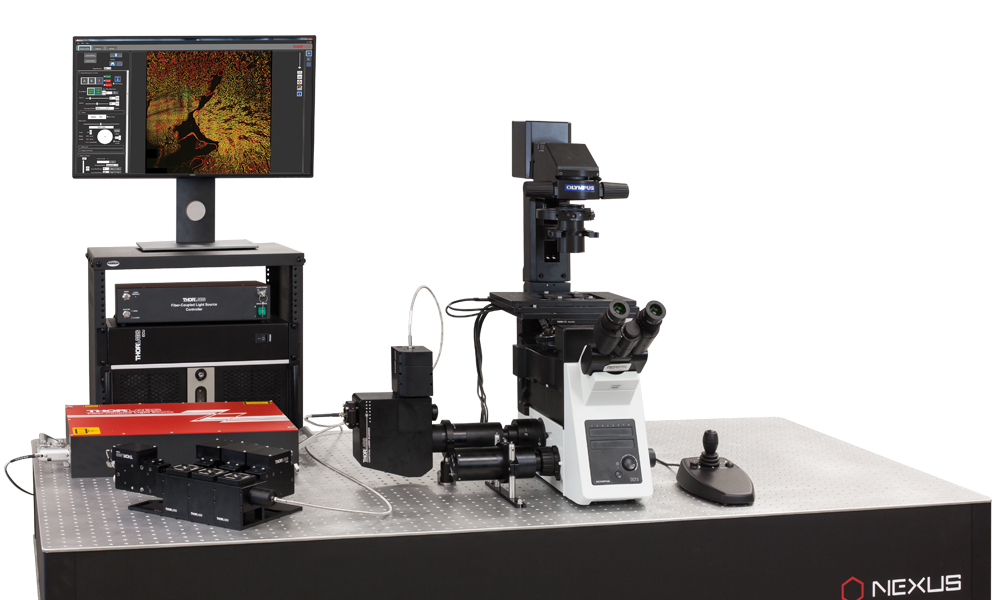
confocal microscopy microscope thorlabs optical system olympus upgrade
1. Introduction The major application of confocal microscopy in the biomedical sciences is. To build an image, the focused spot of light must be scanned across the speci-men in some way. In Minsky's original microscope the beam was stationary and the specimen itself was moved on a vibrating stage.
The confocal microscope is usually operated in epi-fluorescence mode although it can also be used in reflected and transmitted light mode. Most confocals are equipped with one or more lasers that can excite most common fluorescent probes excited by visible light ( FTIC, Rhodamines, Cyanine dyes.)
Confocal microscopy is broadly used to resolve the detailed structure of specific objects within the cell. As a distinctive feature, confocal microscopy enables the creation of sharp images of the exact plane of focus, without any disturbing fluorescent light from the background or other regions of

processes

Confocal microscopy, most frequently confocal laser scanning microscopy (CLSM) or laser confocal scanning microscopy (LCSM)...
A confocal microscope differs from a "classical" optical microscope (see chapter ) in Detailed examination of existing confocal microscope designs is beyond the scope of this chapter. Now let us examine mathematically how and how much the contrast is changed when utilizing the
The principal of confocal microscopy was patented by Marvin Minsky in 1957 but it took a good few years before it was fully developed to incorporate a laser scanning process. The technique essentially scans an object point-by-point using a focused laser beam to allow for a 3-D reconstruction.
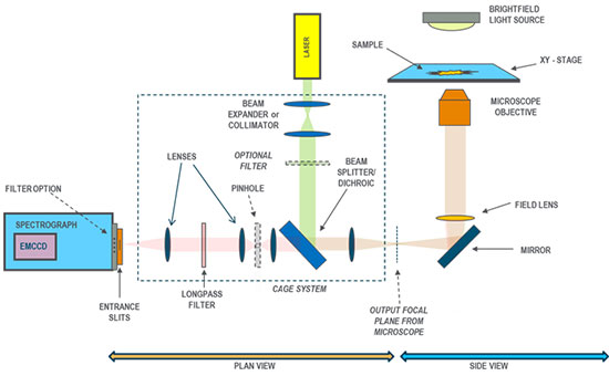
30, 2021 · This surface will act as a dummy surface placeholder for your pinhole. Put 70 mm between the pinhole and the first surface of the collimating optics. Add the surfaces for a collimating lens to the end of the system. In this example, the lens thickness is 6mm.

imaging resolution microscope super software confocal olympus module scanning system laser solution microscopes science
Fundamental Set-up of Fluorescence Microscopes: confocal vs. wideeld. Confocal Fluorescence Microscopy. How many pixels are needed to reproduce the object with the full resolution obtained by the microscope? -> Nyquist criterion for digital resolution: smallest resolved structures
Confocal microscopy is a versatile imaging method through which viewing cells and tissues is made possible. It is a major improvement from conventional light microscopy as it also helps create images in 3D.
Confocal microscopy, most frequently confocal laser scanning microscopy (CLSM), is a powerful technique to produce sharp images of a sample that would otherwise appear blurred when viewed under a conventional microscope. Reconstruction of three-dimensional structures from
confocal spectroscopy solutions microscope fluorescence experiment inverted microspectroscopy modular setup learning configuration overview
It also discusses how to build a merit funciton for optimization and how to use the Convert to NSC Group tool to convert system from Sequential Mode to Non-Sequential Mode. This article will provide a walk-through of the design to show how to acurrately model confocal microscopes in OpticStudio.
Introductory Confocal Concepts. Confocal microscopy offers several advantages over The laser scanning confocal microscope (LSCM) is currently the most widely used confocal variation for biomedical research applications. In order to build an image using the confocal principle,
Confocal microscopy provides only a marginal improvement in both axial (z; along the optical axis) and lateral (x and y; in the specimen plane) optical resolution, but is able to exclude secondary fluorescence in areas removed from the focal plane from resulting images.

confocal microscopy
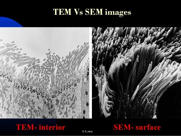
microsopy tem
Confocal microscopes are much more complex than widefield systems. How much does a microscope cost? A lot. A good fluorescence scope system might be about $100k, a confocal more like $400k. Medical Sciences Research Building III Cluster.
Laser Scanning Microscope : Besides other things I'm very interested in lasers, the microscopic world, and to make things visible that aren't visible with normal means. Nothing fancy as a Fluorescent Confocal Laser Scanning Microscope, but a straight forward LSM.
This paper describes how the confocal advantage comes about and how it is implemented in actual A confocal microscope does have slightly higher resolution than a wide eld microscope, but this is not Confocal optical microscopy. 437. Figure 8. p(ζ, ρ) for the focal plane and planes parallel to it: in (a) The main idea, of course, is to move the focused spot over an object, so as to build up an image.

infinity corrected microscope components optical lenses optics physics building objective diagram tube olympus length through field ray finite scratch conjugate
